


Protein concentration is a critical step in many molecular biology and biochemistry workflows, from enzyme purification to antibody production and mass spectrometry sample preparation. An ultrafiltration tube provides a streamlined, reliable method for concentrating protein samples by leveraging size-selective membrane technology and centrifugal force. Understanding the precise mechanism by which an ultrafiltration tube operates allows researchers to optimize concentration protocols, preserve protein integrity, and achieve reproducible results across diverse experimental conditions.
The effectiveness of an ultrafiltration tube in protein concentration stems from its ability to separate molecules based on molecular weight cutoff while maintaining sample stability and minimizing protein loss. This process combines membrane filtration principles with practical laboratory centrifugation, creating a system that removes excess buffer, salts, and small contaminants while retaining target proteins above a defined size threshold. The following sections explain the operational mechanism, design factors, and practical considerations that determine how effectively an ultrafiltration tube concentrates protein samples in real-world applications.
Membrane-Based Size Exclusion Mechanism
Molecular Weight Cutoff Principle
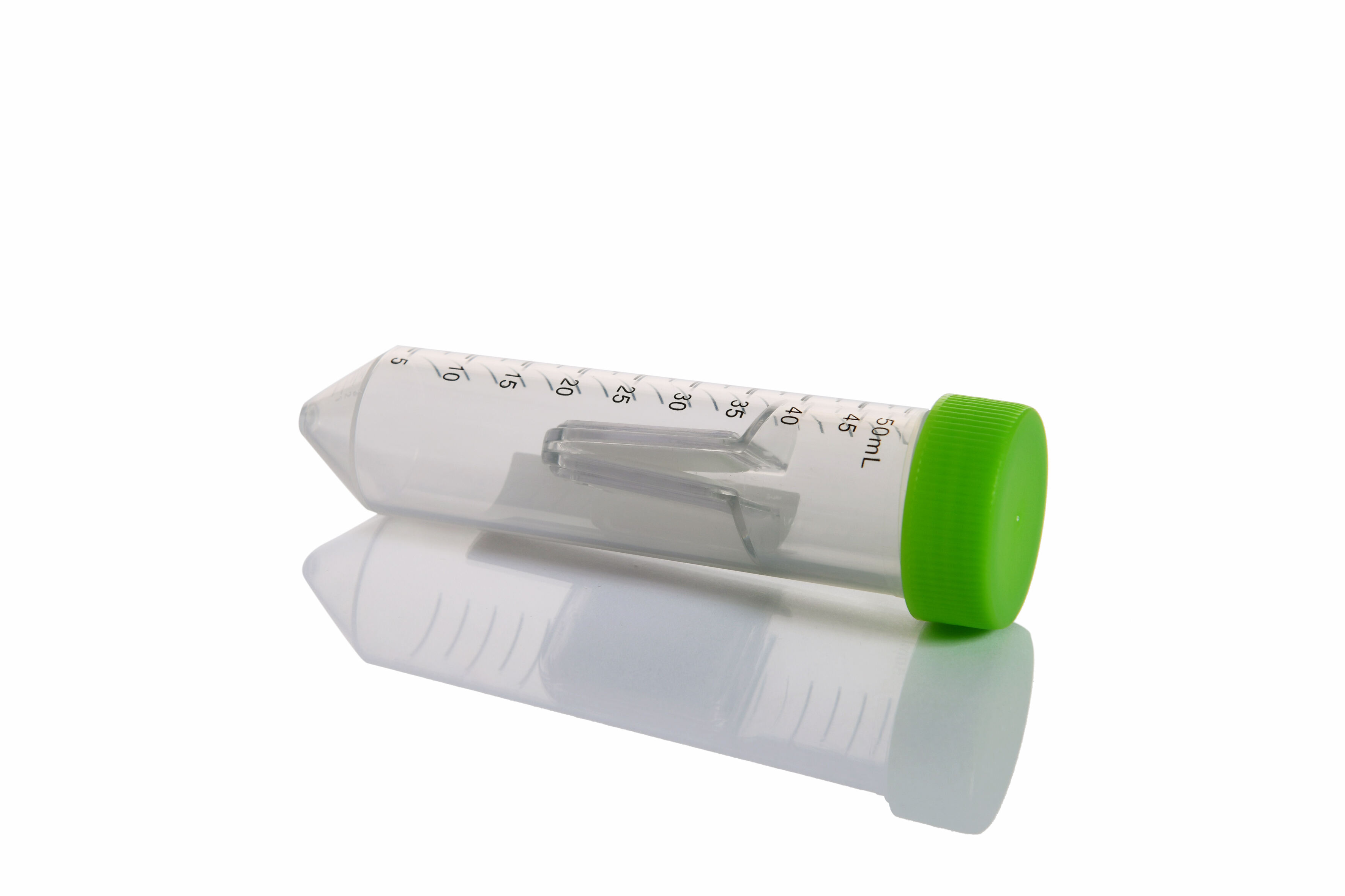
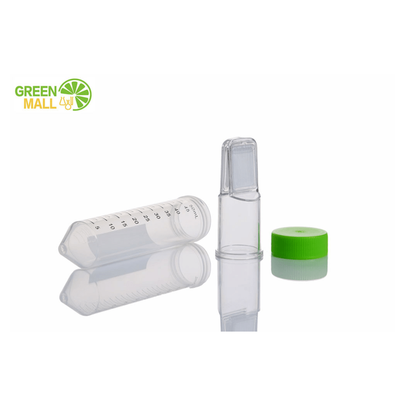
The core operational principle of an ultrafiltration tube relies on a semi-permeable membrane with a defined molecular weight cutoff value, typically ranging from 3 kDa to 100 kDa depending on the target protein size. The membrane functions as a physical barrier that allows water, buffer components, and small molecules below the cutoff threshold to pass through during centrifugation, while retaining larger protein molecules in the upper chamber. This size-selective filtration creates a concentration gradient that drives fluid movement without subjecting proteins to harsh chemical treatments or extreme temperature conditions.
The molecular weight cutoff selection directly influences concentration efficiency and protein recovery rates. When researchers choose an ultrafiltration tube with a cutoff value significantly lower than their target protein molecular weight, retention rates typically exceed 95 percent, ensuring minimal sample loss during the concentration process. Conversely, selecting a cutoff too close to the protein size may result in partial protein passage through the membrane, reducing final yield and compromising experimental outcomes.
The membrane material composition affects both filtration performance and protein compatibility. Most ultrafiltration tube membranes consist of modified polyethersulfone or regenerated cellulose, materials chosen for their low protein binding characteristics and chemical resistance across a wide pH range. These materials maintain structural integrity under centrifugal forces while presenting minimal surface interaction with protein molecules, which helps preserve native protein conformation and biological activity throughout the concentration workflow.
Centrifugal Force Application
Centrifugal force serves as the driving mechanism that propels filtrate through the ultrafiltration tube membrane while retaining concentrated protein in the sample chamber. When the ultrafiltration tube is placed in a standard laboratory centrifuge and spun at specified speeds, typically between 3,000 and 14,000 relative centrifugal force units, hydrostatic pressure builds within the upper chamber, forcing buffer and small molecules through membrane pores into the collection tube below. This process continues until the volume reduction reaches the desired concentration factor or until the sample reaches maximum viscosity limits.
The relationship between centrifugation speed, duration, and concentration efficiency follows predictable patterns that researchers can optimize for specific protein types and starting volumes. Lower centrifugation speeds applied over longer time periods generally produce gentler concentration with reduced risk of protein denaturation, making this approach suitable for sensitive or aggregation-prone proteins. Higher speeds accelerate the concentration process but may increase membrane fouling and protein-membrane interaction, particularly with hydrophobic or charged protein species.
Temperature control during centrifugation significantly impacts protein stability and concentration effectiveness. Most ultrafiltration tube protocols recommend performing centrifugation at four degrees Celsius to minimize protein degradation, reduce microbial growth, and decrease the risk of temperature-induced aggregation. Refrigerated centrifuges equipped with appropriate rotor configurations allow researchers to maintain consistent low temperatures throughout the concentration process, preserving enzymatic activity and structural integrity of temperature-sensitive protein samples.
Design Features That Enhance Concentration Efficiency
Membrane Surface Area Optimization
The effective membrane surface area within an ultrafiltration tube directly correlates with concentration speed and throughput capacity. Larger membrane areas provide more filtration pathways for buffer passage, reducing the time required to achieve target concentration factors and minimizing the duration proteins remain under centrifugal stress. Manufacturers engineer ultrafiltration tube membrane geometries to maximize surface area within compact form factors, often incorporating vertically oriented membrane configurations that increase functional area without expanding overall device dimensions.
The vertical membrane design employed in most ultrafiltration tube models creates a thin liquid layer across the membrane surface during centrifugation, promoting uniform flow distribution and preventing localized concentration gradients that could trigger protein precipitation. This geometry ensures that proteins near the membrane surface experience similar concentration conditions as those in the bulk sample volume, reducing the risk of aggregation hot spots and maintaining sample homogeneity throughout the concentration cycle.
Membrane surface treatment technologies further enhance concentration performance by reducing non-specific protein adsorption. Modern ultrafiltration tube membranes often incorporate hydrophilic surface modifications that create a water layer between the membrane material and protein molecules, minimizing direct protein-membrane contact and improving overall protein recovery. These surface treatments prove particularly valuable when concentrating proteins with exposed hydrophobic patches or those prone to surface-mediated aggregation.
Dead Volume Minimization
Dead volume, defined as the minimum sample volume that remains in the ultrafiltration tube after maximum concentration, represents a critical design parameter affecting overall sample recovery and final concentration factors. High-quality ultrafiltration tube designs minimize dead volume through optimized chamber geometry, allowing researchers to achieve concentration factors of 10 to 100-fold while maintaining practical sample retrievability. Typical dead volumes range from 10 to 50 microliters depending on tube format and membrane area, directly determining the maximum achievable protein concentration.
The relationship between starting sample volume and final concentrated volume determines practical concentration limits for any ultrafiltration tube application. When starting volumes significantly exceed membrane capacity, researchers may need to perform concentration in multiple cycles or select larger-format devices with increased membrane areas and chamber volumes. Conversely, small starting volumes approaching the dead volume threshold may not justify ultrafiltration tube concentration, as alternative methods such as vacuum centrifugation or precipitation might offer better recovery rates.
Chamber geometry design influences both dead volume characteristics and sample recovery efficiency. Conical chamber bottoms concentrate retained sample into minimal volumes while facilitating complete pipette-based recovery, whereas flat-bottom designs may leave residual sample distributed across larger surface areas. The ultrafiltration tube chamber shape selection should align with downstream application requirements, particularly when recovering concentrated proteins for applications requiring precise volume control or minimal dilution.
Factors Affecting Protein Retention and Recovery
Protein Physicochemical Properties
The physicochemical characteristics of target proteins substantially influence retention efficiency and recovery rates during ultrafiltration tube concentration. Protein molecular weight represents the primary determinant of retention, with larger proteins showing near-complete retention when membrane cutoff values are appropriately selected. However, protein shape also affects retention behavior, as elongated or flexible proteins may exhibit smaller effective molecular diameters than globular proteins of identical molecular weight, potentially allowing passage through membrane pores sized based solely on molecular weight calculations.
Protein charge distribution and isoelectric point impact membrane interaction and retention characteristics throughout the concentration process. Proteins carrying net charges similar to membrane surface charges experience electrostatic repulsion that reduces membrane fouling and enhances recovery rates. Conversely, proteins with opposite charge characteristics may exhibit increased membrane binding, particularly near their isoelectric points where reduced electrostatic repulsion allows closer membrane approach and potential adsorptive interactions.
Protein hydrophobicity directly affects membrane binding propensity and recovery efficiency when using ultrafiltration tube systems. Highly hydrophobic proteins or those with significant exposed hydrophobic surface patches show greater tendency toward membrane adsorption, especially on membranes lacking extensive hydrophilic surface modifications. Researchers concentrating hydrophobic proteins may benefit from adding low concentrations of non-ionic detergents or adjusting buffer composition to reduce protein-membrane hydrophobic interactions while maintaining protein solubility and stability.
Buffer Composition and pH Control
Buffer composition exerts significant influence on protein behavior during ultrafiltration tube concentration, affecting protein solubility, membrane interaction, and overall recovery rates. Buffer selection must balance protein stability requirements with membrane compatibility, avoiding components that might promote membrane fouling or alter membrane selectivity characteristics. Common buffer systems including phosphate, Tris, and HEPES generally perform well in ultrafiltration tube applications, provided ionic strength remains within ranges that support protein solubility without inducing excessive osmotic pressure effects.
The pH environment during concentration affects both protein stability and membrane performance characteristics. Operating near the protein isoelectric point increases aggregation risk and may reduce recovery rates due to decreased electrostatic repulsion between protein molecules. Most ultrafiltration tube protocols recommend maintaining pH at least one unit away from the protein isoelectric point, ensuring adequate protein charge that promotes electrostatic stabilization and reduces self-association tendencies during the concentration process.
Glycerol and other viscosity-modifying additives present in protein storage buffers can significantly impact ultrafiltration tube concentration rates and final achievable concentration factors. High glycerol concentrations increase solution viscosity, reducing filtrate flow rates through membrane pores and extending required centrifugation times. When glycerol removal is not essential for downstream applications, researchers may optimize concentration protocols by first performing a buffer exchange into low-viscosity medium using the ultrafiltration tube, then concentrating the exchanged sample to target volume with improved efficiency.
Practical Optimization Strategies
Pre-Concentration Sample Preparation
Sample clarification before loading into an ultrafiltration tube significantly improves concentration efficiency and reduces membrane fouling. Removing particulate matter, cell debris, and aggregated proteins through centrifugation or filtration prevents these materials from accumulating on the membrane surface and blocking filtration pathways. A standard clarification protocol involves centrifuging crude samples at 10,000 to 20,000 relative centrifugal force for 10 to 15 minutes, followed by careful transfer of supernatant to the ultrafiltration tube without disturbing pelleted material.
Protein solubility assessment prior to concentration prevents precipitation-related losses and membrane fouling during the ultrafiltration tube workflow. Researchers should verify that protein remains fully soluble at concentrations significantly exceeding the target final concentration, ideally testing solubility at twice the intended endpoint concentration. When solubility limits approach target concentration values, adjusting buffer composition, adding stabilizing agents, or accepting lower concentration factors may be necessary to maintain protein stability and recovery throughout the concentration process.
Sample volume management relative to ultrafiltration tube capacity optimizes concentration efficiency and reduces processing time. Loading the maximum recommended sample volume for a given ultrafiltration tube format minimizes the number of concentration cycles required while maintaining appropriate membrane area-to-sample volume ratios. For large starting volumes, selecting higher-capacity ultrafiltration tube formats or performing sequential concentration steps with volume consolidation between stages provides more efficient pathways to target concentration than attempting to process excessive volumes in undersized devices.
Process Monitoring and Endpoint Determination
Monitoring concentration progress during ultrafiltration tube processing prevents over-concentration and allows timely intervention if unexpected issues arise. Periodic volume checks during extended centrifugation runs enable researchers to track concentration rates and estimate remaining processing time. Visual inspection of the sample chamber provides immediate feedback regarding sample appearance, detecting early signs of precipitation or unusual viscosity increases that might indicate approaching solubility limits or protein aggregation.
Determining optimal concentration endpoints requires balancing the desire for maximum volume reduction against practical constraints of protein solubility, sample viscosity, and recovery efficiency. Pushing concentration beyond protein solubility limits triggers precipitation and irreversible sample loss, while excessive viscosity increases in the sample chamber can slow filtration rates to impractical levels and complicate accurate pipetting during sample recovery. Most successful ultrafiltration tube protocols target concentration factors that maintain protein concentrations at 60 to 80 percent of known solubility limits, providing safety margins that account for local concentration variations near the membrane surface.
Recovery technique optimization ensures maximum transfer of concentrated protein from the ultrafiltration tube sample chamber to collection vessels. Rinsing the sample chamber with small volumes of appropriate buffer captures residual protein adhering to chamber walls and membrane surfaces, typically improving overall recovery by 5 to 15 percent. Multiple gentle rinses using small buffer volumes prove more effective than single large-volume washes, as they maintain higher protein concentrations during recovery and reduce total dilution of the concentrated sample.
Troubleshooting Common Concentration Challenges
Slow Filtration Rates
Unexpectedly slow filtration rates during ultrafiltration tube concentration often indicate membrane fouling, excessive sample viscosity, or inappropriate centrifugation parameters. Membrane fouling occurs when proteins, aggregates, or particulates accumulate on the membrane surface, blocking pores and restricting buffer flow. Addressing fouling typically requires improved sample clarification before loading, selection of membranes with low protein binding characteristics, or adjustment of buffer composition to reduce protein-membrane interactions.
High sample viscosity naturally slows filtration rates by increasing resistance to fluid flow through membrane pores. Viscosity effects become particularly pronounced when concentrating proteins to high final concentrations or when working with naturally viscous samples such as antibody preparations or glycoprotein solutions. Managing viscosity-limited concentration may require accepting lower final concentration factors, increasing centrifugation speeds within membrane specifications, or performing buffer exchange to remove viscosity-enhancing components before final concentration.
Incorrect centrifugation speed or rotor selection can substantially limit filtration efficiency in ultrafiltration tube applications. Operating below manufacturer-recommended speeds reduces the hydrostatic pressure driving filtration, unnecessarily extending processing times. Using fixed-angle rotors instead of swinging-bucket designs may alter the effective membrane orientation during centrifugation, potentially reducing filtration efficiency for some ultrafiltration tube designs optimized for specific rotor configurations.
Protein Loss and Recovery Issues
Lower than expected protein recovery from ultrafiltration tube concentration commonly results from membrane adsorption, protein aggregation, or passage through the membrane due to inappropriate cutoff selection. Membrane adsorption losses typically affect hydrophobic proteins or those with charge complementarity to membrane surfaces, with losses ranging from 5 to 30 percent depending on protein characteristics and membrane type. Minimizing adsorption requires selecting membranes with extensive hydrophilic modifications, adding low concentrations of non-ionic detergents, or including carrier proteins that compete for membrane binding sites.
Protein aggregation during concentration leads to both functional sample loss and potential membrane fouling that further reduces recovery of remaining soluble protein. Aggregation risk increases with protein concentration, making it particularly problematic during the final stages of ultrafiltration tube processing when local protein concentrations near the membrane surface can exceed bulk solution values. Preventing aggregation requires careful buffer optimization, temperature control, and recognition of protein-specific concentration limits beyond which aggregation becomes thermodynamically favorable.
Protein passage through the membrane despite appropriate molecular weight cutoff selection may occur with elongated proteins, multi-domain proteins connected by flexible linkers, or partially denatured proteins with altered hydrodynamic properties. When passage losses exceed 10 percent, researchers should verify protein integrity through analytical methods, consider selecting ultrafiltration tube membranes with lower cutoff values, or explore alternative concentration methods better suited to proteins with unusual structural characteristics or conformational flexibility.
FAQ
What concentration factor can typically be achieved with an ultrafiltration tube?
Most ultrafiltration tube systems routinely achieve concentration factors between 10-fold and 50-fold, with some applications reaching 100-fold concentration depending on starting volume, protein characteristics, and device dead volume. The practical upper limit is determined by protein solubility, sample viscosity at high concentration, and the minimum retrievable volume specific to the ultrafiltration tube design being used.
How long does protein concentration typically take using an ultrafiltration tube?
Concentration time varies from 15 minutes to several hours depending on starting volume, target concentration factor, protein properties, and centrifugation speed. A typical 500 microliter sample concentrated 10-fold using an ultrafiltration tube with 10 kDa cutoff requires approximately 30 to 60 minutes at 14,000 relative centrifugal force under optimal conditions with dilute protein solutions in low-viscosity buffers.
Can ultrafiltration tubes be reused for multiple protein concentration cycles?
Ultrafiltration tubes are generally designed as single-use devices to prevent cross-contamination and ensure consistent performance. While membrane cleaning and regeneration protocols exist, they cannot guarantee complete removal of all bound proteins or restoration of original membrane properties. The risk of sample contamination and reduced filtration efficiency makes reuse inadvisable for most research applications requiring reproducible results.
What should I do if my protein precipitates during ultrafiltration tube concentration?
If precipitation occurs during concentration, immediately stop centrifugation and attempt to redissolve precipitated protein by diluting with appropriate buffer while gently mixing. For future attempts, reduce the target concentration factor, optimize buffer composition by adding stabilizing agents or adjusting pH and ionic strength, perform concentration at lower temperature, or consider alternative concentration methods such as precipitation-based approaches followed by controlled resolubilization in minimal volumes.
Table of Contents
- Membrane-Based Size Exclusion Mechanism
- Design Features That Enhance Concentration Efficiency
- Factors Affecting Protein Retention and Recovery
- Practical Optimization Strategies
- Troubleshooting Common Concentration Challenges
-
FAQ
- What concentration factor can typically be achieved with an ultrafiltration tube?
- How long does protein concentration typically take using an ultrafiltration tube?
- Can ultrafiltration tubes be reused for multiple protein concentration cycles?
- What should I do if my protein precipitates during ultrafiltration tube concentration?